Library search
Search Results
-
 Optimization of surgical treatment of chronic antero-medial dislocations of the radial head in children
Catalog of dissertations and abstractsThe aim of the research work is to improve the results of surgical treatment of chronic anterior-medial dislocations of the radial head in children based on the improvement of the method of surgical treatment.
The object of the study was 83 patients with chronic antero-medial dislocations of the head of the radius, treated in the department of the consequences of childhood injuries of the Samarkand branch of the RSSPMC of Traumatology and Orthopedics for the period 2017-2020. The scientific novelty of the research work is the following: it is proved by histological examination that, in case of injuries from up to 1 month ago, the anterior wall of the joint capsule is thin and elastic, which is easily stretched, and from 3 months ago, it thickens, scars and forms fibrous tissue; the possibility of using a fibrous-modified joint capsule for annular ligament plasty in the surgical treatment of chronic antero-medial dislocations of the radial head from 3 months ago was proved; the tactics of surgical treatment of chronic antero-medial dislocations of the head of the radius depending on the deformity of the bones of the forearm were determined; a direct relationship between the results of surgical treatment of chronic antero-medial dislocations of the head of the radius, depending on the duration of the injury, has been proven.
The introduction of research results. Based on the obtained scientific results on the optimization of surgical treatment of chronic antero-medial dislocations of the radial head in children:
based on the results of the development of a method for annular ligament plasty, a patent for an invention was obtained from the Intellectual Property Agency of the Russian Federation “A method for the surgical treatment of chronic anterior medial dislocation of the radial head in children by capsuloplasty according” (patent № 2749870 dated 06/17/2021). The results obtained made it possible to improve the results of surgical treatment, to reduce the period of penetration in the hospital and after the surgical rehabilitation period; based on the results of scientific research on the surgical treatment of chronic anterior medial dislocations of the radial head, the guidelines “Surgical treatment of chronic dislocations of the radial head in children” were approved (Conclusion of the Ministry of Health of the Republic of Uzbekistan 8 n-z / 81 dated February 21, 2022). The results obtained have improved the quality of early diagnosis and treatment of patients with chronic anterior medial dislocations of the radial head in children; based on the results of scientific research on the surgical treatment of chronic antero-medial dislocations of the head of the radius, the methodological recommendations “Conclusion of the Ministry of Health of the Republic of Uzbekistan 8 n-z / 289 of August 31, 2021” were approved. The results obtained have improved the quality of early diagnosis and treatment of patients with chronic anterior medial dislocations of the radial head in children;
Scientific results have been introduced into the practice of healthcare (Conclusion of the Ministry of Health 08-32955 of October 24, 2022), in particular, the Samarkand branch of the Republican Specialized Scientific and Practical Medical Center for Traumatology and Orthopedics, the Bukhara branch of the Republican Scientific Center for Emergency Medical Care, and the Samarkand Regional Children's Multidisciplinary Medical Center. The proposed method for the treatment of chronic anterior-medial dislocations of the radial head in children allowed to reduce the frequency of relapses, increase excellent and good results from 75.6% to 92.9%.
The structure and scope of the dissertation. The dissertation consists of an introduction, 5 chapters, conclusion, conclusions, practical recommendations, a list of references and applications. The volume of the dissertation is 109 pages. -
 Surgery engrained anteromedial dislocation of the radial head in childrenPresented results of surgical treatment of old dislocations of caput of radii. Findings of searching were based on results of surgical treatment 37 patients with dislocations of caput radii on different reasons. Patients were done open reduction of caput of radii with repair of ligament annular. For modernization methods of re-pair of ligament annular we used its capsule of the joint and pats of ligament annular which has more anato-my-physiologically for elbow joint. By this method we have good and satisfactory functional results on 97,4% cases and anatomical on 95,7% cases
Journal problems of biology and medicine -
 Improvement of surgical aspects in providing help for sufficients wi thcombined pelvic and femoral bone damages
Catalog of dissertations and abstractsThe aim of the research work is to improve the results of treatment of patients with combined injuries of the pelvis and femur, by developing tactical and technical aspects based on the severity of the injury and the severity of the condition.
The object of the study was 130 patients with injuries of the pelvic and hip bones with concomitant trauma, treated at the Republican Scientific Center for Emergency Medical Aid and its Samarkand branch for the period 2016-2021 years.
The scientific novelty of the research work is the following: the structure and frequency of combined injuries of the pelvis and femur in the general structure of injuries, in the structure of injuries to the pelvis and femur separately were evaluated. the risk factors for the development of unsatisfactory results of treatment of concomitant injuries of the pelvis and hip, based on traditional clinical and diagnostic standards, have been determined; a direct relationship has been proven in the dynamics of the condition of the victims and the prognosis, taking into account the type and nature of segmental injuries; the device for external fixation for stable functional minimally invasive osteosynthesis has been improved and the possibility of expanding the indications for surgical treatment for combined injuries of the pelvis and hip in the early period of traumatic disease has been proved; the technical advantages of a complete set of an improved rod device for external fixation have been proved, the pelvic and femoral versions of which make it possible to use them for effective stabilization of the pelvis and hip separately during anti-shock measures, and for the final reposition of bone fragments; the direct dependence of treatment results on the proposed tactics of providing trauma care at an early hospital stage, depending on the type, nature, severity of pelvic and hip injuries, and the severity of the condition has been proved.
The introduction of research results. Based on the results of scientific research to improve the surgical aspects of providing assistance to victims with concomitant injuries of the pelvis and femur: based on the results of the development of a device for the treatment of fractures, a patent for an invention was obtained from the Intellectual Property Agency of the Russian Federation "Apparatus for the treatment of combined fractures of the pelvic and hip bones" (patent No. 2749897 dated 06/18/2020). The results obtained made it possible to improve the tactics of surgical treatment of patients, to shorten the period of hospitalization and the period of postoperative rehabilitation, to ensure the possibility of patients with minimal economic costs; on the basis of the results of scientific research on the diagnosis and treatment of concomitant injuries of the pelvic and femur bones, methodological recommendations were approved "Method for the treatment of victims with concomitant injuries of the pelvis and hip, depending on the severity" (Conclusion of the Ministry of Health of the Republic of Uzbekistan No. 8 n-z / 288 dated August 31, 2021 of the year). The results obtained made it possible to improve the quality of wound diagnosis and rehabilitation of patients with injuries of the pelvic and hip bones in concomitant injury; approved methodological recommendations "Tactics of rendering assistance to victims with combined injuries of the pelvis and hip, taking into account the severity of the condition" (Conclusion of the Ministry of Health of the Republic of Uzbekistan No. 8 n-z / 288 of August 31, 2020). The results obtained made it possible to improve the tactical and technical aspects in the treatment of injuries to the pelvic and hip bones, based on the severity of the injury and the severity of the patient's condition.
Scientific results have been introduced into the practical activities of healthcare, in particular, the Samarkand branch of the Republican Specialized Scientific and Practical Medical Center of Traumatology and Orthopedics, the Jizzakh Branch of the Republican Scientific Center for Emergency Medical Aid, the Samarkand branch of the Republican Scientific Center for Emergency Medical Aid (certificate of the Ministry of Health No. 08-09 / 18979 dated December 02, 2021). The proposed tactics for the treatment of combined injuries of the pelvis and femur made it possible to reduce the incidence of postoperative complications of excellent and good long-term functional results from 66.1% to 92.6%.
The structure and scope of the dissertation. The dissertation consists of an introduction, five chapters, conclusions, practical recommendations, a list of referencesand applications. The volume of the text material of the work is 111 pages.
-
 EXPERIENCE OF VASCULARIZED AND NONVASCULARIZED AUTOTRANSPLANTS USE FOR LOWER JAW DEFECTS SUBSTITUTION AFTER ITS RESECTIONSummary. To 43 patients after lower jaw resection with disarticulation (on the occasion of neoplasm) the defects werereplaced by vascularized autotransplants of the 2nd radius of pedis and vascularized fibula autotransplants in combination with titanium implants, hi all cases positive results were received. The authors consider that microsurgery' with vascularized fibula autotransplants in combination with titanium implants was an optimal way to replace lower jaw defects after its resection.
Medicine and innovations -
 Ultrasound assessment of varying degrees of hip dysplasia in neonatesCongenital hip disorders such as hip dysplasia, hip subluxation, and hip dislocation are among the most common musculoskeletal disorders, accounting for up to 15% of orthopaedic pathology, leading to statico-dynamic disturbances and limitation of vital functions and early disability. The incidence of these diseases according to different authors ranges from 5.3 to 50 and even 200 cases per 1000 newborns. Treatment initiated in the first month of a child's life is most effective and leads to complete anatomical and functional restoration of the joint in 88% to 98% of cases. In cases of late treatment, 10-60% of children develop one of the severe diseases - dysplastic coxarthrosis, aseptic necrosis of the femoral head, compensatory scoliosis of the spine or other anatomical and functional changes.
Journal of hepato-gastroenterological research -
 Surgical treatment of non-consolidated fractures of the humeral condyle head in childrenThe experience of operative treatment in 116 children with incomplete fractures and pseudoarthrosis of the condyle of the humeral head is presented. The reasons for nonunion are errors of diagnosis and treatment of patients. Also important is the intraarticular nature of the fracture, the condition of the broken fragment from cartilage tissue and bone tissue, the contact of heterogeneous tissues in the presence of a lateral proximal small (4-5mm) displacement of the bone fragment, which is the cause of the extended period of bone-cartilage callus beyond the redistribution the accepted period of immobilization of the joint. A method for fixing a bone fragment, proposed by the authors, and a method of operation for treating pseudoarthrosis of the humeral condyle of the humerus by bone grafting, taken from the diaphysis of the fibula of the patient himself, is described. Excellent results were obtained in 43.3%. good results were observed in 46.2%, satisfactory - in 5.3%, and unsatisfactory - in 5.3% of patients.
Doctor's Herald -
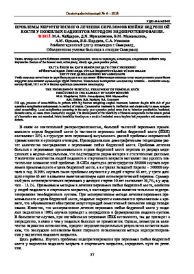
 Одним из важных вопросов современной травматологии являются лечение и профилактика последствий переломов проксимального конца бедренной кости. Среди длинных костей самая прочная - бедренная. Переломы бедренной кости состав- люет 6,5-10,5% от общего количества переломов костей [5]. Переломы проксимального конца бедренной кости составляют от 6 до 18% от всех переломов бедренной кости
 -
 Морфология клеток диффузной эндокринной системы тонкой кишки животных с сенильным остеопорозомЦель исследования. Изучение морфологических и морфометрических показателей клеток диффузной эндокринной системы тонкой кишки и степени минерализации бедренной кости у кроликов и морских свинок старческого возраста. Материал и методы исследования. Изучена тонкая кишка у 5 крольчих 6-7-летнего возраста и у 6 морских свинок-самок 5-6-летнего возраста. Криостатные срезы из нефиксированного материала обработаны раствором глиоксиловой кислоты, парафиновые срезы окрашены гематоксилином и эозином, по методу Ван- Гизона и импрегнированы азотнокислым серебром по Гримелиусу. Степень минерализации бедренной кости определяли сравнением веса золы после сжигания ее к первоначальному весу костей в процентах. Плотность распределения эндокринных клеток определяли точечным методом на стандартной площади. Результаты. Установлено достоверное уменьшение числа (плотность распределения) эндокринных клеток слизистой оболочки и снижение степени минерализации бедренной кости у старых крольчих и морских свинок - самок. Обнаружены дегенеративные изменения некоторых эндокринных клеток в виде вакуолизации их цитоплазмы и снижения степени флюоресценции эндокринных клеток открытого типа старых лабораторных животных. Заключение. В стенке тонкой кишки старых крольчих и морских свинок - самок происходит уменьшение плотности распределения, снижение степени флюоресценции эндокриноцитов и степени минерализации бедренной кости. Отмечается параллелизм между изменением числа эндокринных клеток стенки тонкой кишки и минерализацией бедренной кости.
Doctor's Herald -
 The results of the application of compressesive-distractive osteosynthesis at refract tibia diaphysisThere results of osteosynthesis of tibia by Ilizarov in 540 patients are mentioned in the article. Patients were examined by dividing them into 2 groups. In 486 patients who were in the first group, it was tried to insert broken bones were at once, they were attached to the Ilizarov installation consisting of 4 rings. The part of bones that was impossible to insert at once, were inserteded gradually with the help of the installation. In 80% of patients the bones grew together in time. In 20% there was a slow fusion not fusion and false joints. In 54 patients who were in the second group, the bones were inserted at the same time with the help of IIT, and are attached to the installation of the Ilizarov. In all Patients who were in the second group but one (he had con- comitant diseases, obesity -III degree, diabetes) bones have grown together during. The reason of the fact that the bones were inserted at the same time and were strongly attached to the seam.
Journal problems of biology and medicine -
 Sonography and magnetic resonance imaging of the hand joints in rheumatoid arthritis
Catalog of abstractsSubject of the inquiry: 116 patients with rheumatoid arthritis of the hand joints and 25 healthy subjects.
Aim of the inquiry: improvement of the diagnosis of rheumatoid arthritis of the hand joints using sonography and magnetic resonance imaging.
Methods of inquiry: X-ray, sonography, magnetic resonance imaging and magnetic resonance imaging with contrast enhancement.
The results achieved and their novelty: For the first time data were presented on the role of sonography, magnetic resonance imaging and magnetic resonance imaging with contrast enhancement in the diagnosis of rheumatoid arthritis of the hand joints. The findings have shown that sonography in hand joint rheumatoid arthritis allowed detection of changes in soft tissues, synovial capsule, joint surfaces and ligaments. Diagnostic value was given of sonography in revealing characteristic sonographic signs of rheumatoid arthritis. Magnetic resonance imaging was highly informative radiological method to detect synovitis, changes of synovial capsule and subchondral cysts. Magnetic resonance imaging with contrast enhancement reliably detected the degree of activity and severity of the rheumatoid process.
Practical value: consisted in revealing and describing characteristic sonographic and MRI signs of hand joint rheumatoid arthritis and in the developed radiological algorithm of the disease.
Degree of embed: the results of the investigation were introduced in the practice of rheumatology and radiology departments of the First Tashkent Medical Institute, in teaching process of the Radiology Chair of the First Tashkent Medical Institute.
Sphere of usage: radiology, rheumatology, traumatology and orthopedics. -
 Показатели рентгеновской денситометрии у детей с асептическим некрозом головки бедренной кости
Современная медицина глазами молодых ученыхВ развитии асептического некроза головки бедренной кости большую роль играет совокупность факторов, включающие в себя как нсйрососудистыс нарушения, особый гормональный фон, влияние окружающей среды, так и особенности строения тазобедренного сустава в биомеханическом плане. Процесс перестройки, лежащий в основе любых изменений формы и строения кости, зависит не только от состояния кровоснабжения, но и от условий функциональной нагрузки. Эти два фактора совместно приводят к активизации процессов перестройки кости.
-
 Along with the assessment of the clinical status, the use of radiography of the lungs is significant. The features of the X-ray symptoms of pneumonia in young children were studied depending on the gestational age at birth. In case of prematurity, radiological signs characteristic of continuing immaturity of the lung tissue were revealed, namely, moderate hypoventilation of the lungs, widespread small-focal shadows, limited reticular deformity of the pulmonary pattern and the symptom of “air bronchogram”. From 1 year to 3 years, the above symptoms are somewhat less pronounced, which is consistent with the available information on the response from the interstitial tissue in the age dynamics. Long-term signs of immaturity of lung tissue in children born prematurely leave an imprint on the process of further development of respiratory tract diseases in young children and do not exclude the possibility of age-related predisposition to interstitial lung diseases.
 -
 НАШ ОПЫТ ХИРУРГИЧЕСКОГО ЛЕЧЕНИЯ ВРОЖДЕННОГО ВОЗВЫШЕНИЯ ЛОПАТКИ У ДЕТЕЙ РАННЕГО ВОЗРАСТАВрожденная высокая деформация лопатки, широко известная как деформация Шпренгеля, редкая врожденная деформация неизвестной этиологии возникает из-за прерывания каудальной миграции лопатки в раннее развитие плода, приводящее к приподнятой и гипоплазированной лопатке. Оставленная без лечения деформация Шпренгеля часто приводит к функциональным и косметическим нарушениям, таким как ограничение движений плеча и выраженная ипсилатеральная перепонка шеи. В данной работе мы представляем несколько клинических случаев лечения детей с деформацией Шпренгеля, где рекомендовано при планировании хирургической коррекции врожденного высокого стояния лопаточной кости необходимо учитывать высоту расположения лопатки, сопутствующие изменения в костях грудной клетки, позвоночника, патологические изменения формы и размеров лопаточной кости. А при наличии деформации формы лопаточной кости после полной мобилизации лопатки целесообразно полная коррекция патологической формы для достижения равномерного прилегания ее к поверхности грудной клетки и равномерного скольжения при движениях верхней конечности.
Medicine and innovations -
 Переломы костей голени составляют около 10% всех переломов костей. Выделяют переломы мыщелков большеберцовой кости, диафизов костей голени, лодыжек, переломы головки малоберцовой кости, отрывные переломы бугристости большеберцовой кости. Переломы дистального и проксимального концов костей голени являются внутри- и околосуставными
 -
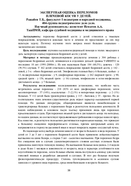
Переломы бедренной кости у детей относятся к тяжелым повреждениям, встречаются довольно часто и лечение их представляет до сих пор нелегкую задачу. Поэтому изучение этого вопроса имеет большое социально -экономические значение.
-
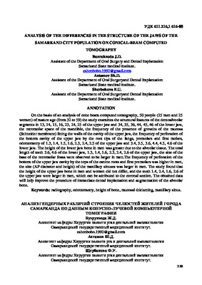 ANALYSIS OF THE DIFFERENCES IN THE STRUCTURE OF THE JAWS OF THE SAMARKAND CITY POPULATION ON CONICAL-BEAM COMPUTEDOn the basis of an analysis of cone beam computed tomography, 50 people (25 men and 25 women) of mature age (from 32 to 59) the study examines the structural features of the dentoalveolar segments in 13, 14, 15, 16, 23, 24, 25 of the upper jaw and 34, 35, 36, 44, 45, 46 of the lower jaw, the retromolar space of the mandible, the frequency of the presence of growths of the mucosa (Schneider membrane) lining the walls of the cavity of the upper jaw, the frequency of perforation of the bottom cavity of the upper jaw by the root tips of the fangs, premolars and first molars, odontometry of 1.3, 1.4, 1.5, 1.6, 2.3, 2.4, 2.5 of the upper jaw and 3.4, 3.5, 3.6, 4.4, 4.5, 4.6 of the lower jaw. The height of the lower jaw bone in men was greater due to the alveolar tissue. The total length of teeth 3.6, 4.6 of the lower jaw, 1.3, 1.4, 1.6, 2.3, 2.4, 2.6 of the upper jaw, the size of the base of the retromolar fossa were observed to be larger in men.The frequency of perforation of the bottom of the upper jaw cavity by the tops of the canine roots and first premolars was higher in men, the size (AP diameter and height) of the maxillary sinuses was larger in men. The study found that the height of the upper jaw bone in men and women did not differ, and the teeth 1.4, 2.4, 1.6, 2.6 of the upper jaw were larger in men, which can be attributed to the coronal section. The obtained data will help improve the procedure of immediate dental implantation and augmentation of the alveolar bone.
Medicine and innovations -
 Воздействия биоактивных покрытий на остинтеграцию имплантов
Topical issues of surgical dentistry and dental implantologyДизайн поверхности имплантатов эволюционировал для решения проблем реабилитации полости рта как в здоровой, так и в нарушенной кости. Например, чтобы победить наиболее распространенные осложнения, связанные с зубными имплантатами, периимплантит и последующую потерю имплантата, поверхности имплантатов были модифицированы, чтобы придать зубному имплантату желаемые свойства и тем самым повысить процент успешного приживления имплантата и расширить показания к их применению. Биоактивные покрытия имеют потенциал для улучшения костной интеграции механически нагруженных ортопедических керамических имплантатов. Есть гипотеза о том, что биоактивное покрытие будет способствовать интеграции отменной кости. Биоактивное покрытие замедлить скорость коррозии сплавов и ускорить процесс заживления кости. Основная Цель биоактивных покрытий - усилить прямое прикрепление живых тканей и тем самым способствовать остеокондукции.
-
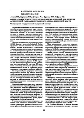 Оценка эффективности органосохранных операций при лечении остеогенной саркомы дистального отдела бедренной костиУвеличение продолжительности и улучшение качества жизни больных остеогенной саркомой дистального отдела бедрен- ной кости T2N0M0 с помощью выбора оптимальной и адекватной тактики хирургического лечения
Journal problems of biology and medicine -
 The problems of surgical treatment of femoral neck fractures in the elderly by endopronesisOld age, presence of comorbidities in patients with hip fracture weighting surgical treatment, however despite with risk of post operation complication cndoproncsis is method of choice. Conservative treatment is ineffective and shown only in cases compensated comorbidity. Local complication occurring in the early post operative period were due to behavior peculiarities burdened. Somatic status, all cases were successfully cropped. The development of the instability of femoral components in the remote period of observation was not recorded. Pelvis instability develops as a result of technical errors implant bad preparation and cementation defects.
Doctor's Herald -
 REHABILITATION OF PATIENTS AFTER ENDOPROSHETICS OF THE HIP JOINT IN ASEPTIC NECROSIS OF THE FEMORAL HEADWe have carried out the rehabilitation of all 64 patients with total arthroplasty from 2015 to 2021, surgery for aseptic necrosis of the femoral head. After EP, the hip joint was divided into two stages: Of these, from the moment of surgery to 3 weeks, this is the early stage. From 3 weeks to 10 weeks - late stage. It is necessary to efficiently perform tasks and exercises for rehabilitation after EPHT by a surgeon and a rehabilitation therapist. At the sanatorium-resort stage, every year patients get physical therapy and physiotherapy for 3 years in a sanatorium-resort environment. Before and after the operation, the difference in the number of movements in 10 seconds was assessed. When performing the test with the operated and unoperated leg, the index of hip abduction increased the most after the operation. The data of the coordination test showed that on the 14th day, it is necessary to productively perform tasks and exercises for rehabilitation after EP while the indicator on the operated leg in the main group was equal to 12.2 m, and in the control group - 11.2 m of movement, which is 25.3% better than the control.
Medicine and innovations -
 Digital method of radiation diagnostics in the assessment of bone structure during dental implantation in patients with diabetes mellitusResults digital method of radiodiagnosis in assessment of bone structure at the dental implantation at patients with diabetes and of intrabone dental miplants osteointegration after restoration of an atrophy of an alveolar bone and dental implantation are shown on a material of 220 patients. Patients with the second type of diabetes at a stage of compensation are in comparative group. For the analysis of results of osteointegration and stability miplants we were used the device «Osstell ISQ». it was manufactured by firm Integration Diagnostics (Sweden), it defining frequency-resonant analysis and stability factor of implants by method RFA (Resonance Frequency Analysis), which registries of resonant electromagnetic fluctuations of implants and a surrounding bone influences on them with electromagnetic field. ISQ (Implant stability Quotient) is expressed on a scale from one to hundred. The obtained data allows assuming influence of objective methods of research of osteointegration dental implants with the subsequent reduction of terms of orthopedic treatment and choice of optimum term of prosthetics. The general average value of implants stability is about 70 units ISQ.
Stomatologiya -
 The role of magnetic resonance imaging in the comprehensive radial diagnosis of volumetric masses of the eye organ
Catalog of abstractsRelevance of the problem. The difficulties of diagnostics of orbital diseases are well known. Especially difficult is intraspecies differentiation among the multitude of tumour, pseudotumour, inflammatory, vascular, endocrine and other diseases occurring here, manifested by the symptom complex of unilateral exophthalmos [Beradze I.N., 1978; Brovkina A.F., 1993].
Malignant intraocular neoplasms are the main cause of death of patients with diseases of the organ of vision, with 45-48% of patients dying from metastases in the first 5 years after enucleation [Alekseeva I.B., 1990, Barkhash S.A.1978, Brovkina A.F..1991, 1997; Keizer R.W.. Viclvoyc G.L.,1986],
Retinoblastoma is the most frequent malignant neoplasm in children. According to different authors, the frequency of its occurrence is 1 case per 14000 - 35000 newborns. [Bobrova N.F. and Vit V.V., 1993; Brovkina A.F., 1997; Provenzale J.M., et al., 1995; Skulski M., et al., 1997; Weber A.L., Mafee M.F, 1992; Wilms G., et al., 1989]. The frequency of patients with the most malignant intraocular tumour in adults - uveal melanoma has recently reached 7-9 people per 1 million population [Brovkina A.F., 1997; Kotslyansky E.O., 1989; Yushko N.A., Peskova L.I., Kalenich L.A., 1989; Peyster R.G., Augsburger J..I., Shields J.A., 1988; Romani A.. Baldeschi L., ct al 1998; Scott I.U., 1998].
The fundamental difference in treatment tactics, depending on the stage of development, size and topography of the tumour, as well as the seriousness of the prognosis in retinoblastomas and melanomas sharply increase the requirements for the accuracy of their differential diagnosis. At the same time, the number of diagnostic errors in ocular tumours continues to be 10-30% even when complex clinical and instrumental examination is applied in specialised ophthalmological centres [Ternovoy S.K., Panfilova G.V., Rogozhin V.A., 1979; Friedman F.E., Malyuta G.D., Kodzov M.V., 1995; Song G.X., 1991].
Widely used in ophthalmological practice traditional diagnostic methods (ophthalmoscopy, gonioscopy, diaphanoscopy, fluorescence angiography, laboratory tests) are insufficient to obtain comprehensive information about the localisation, nature of growth and prevalence of volumetric pathological formations of the eye and orbit. This circumstance and not quite satisfactory results of surgical treatment are the causes of high mortality of patients [Muratova T.T., Nigmanova N.H., Kozlovskaya G.M.. 1989, Naches A.I., 1980; Cheremisin V.M., Trufanov G.E., Kholin A.V., 1991]. Untimely or erroneous recognition of pathological processes of the orbit leads to a sharp deterioration of visual functions, up to blindness, and in some cases to the death of the patient [Yuzhakov A.M., Travkin A.G., Kiseleva O.A., 1991]. All this determines the importance of timely and accurate diagnosis of diseases of the orbit, on the one hand, and the difficulty of such diagnosis - on the other [Gabunia R.I., Kolesnikova E.K., Tumanov L.B., 1982].
The fact that the orbit is closed from direct inspection and palpation by bone walls and the eyeball, indicates the advantage of radial diagnostics in comparison with other methods of examination. In the arsenal of clinicians there is a great variety of methods of clinical-radial diagnostics of orbital pathology, however, at present the information in the literature about their resolving capabilities and significance in comparative aspect is incomplete and not fully studied. The priority of using one or another instrumental investigation, their sequence and expedient combination have not been determined yet. This makes it difficult to choose the optimal standardised approach for diagnosis and adequate treatment [Cheremisin V.M., Trufanov G.E., 1993, Weber A.L., Sabates N.R., 1996; Wenig V.M., Mafee M.F., 1998].
Thus, the study of these and other questions, contributing to the improvement of diagnostics and treatment of patients with neoplasms of the eye and ocular cavity, should be recognised as urgent urgent.
Purpose of the study. Comparative evaluation of magnetic resonance tomography capabilities and development of algorithms for complex radial diagnostics of volumetric formations of the visual organ. To solve this goal we set the following tasks.
1. To study the normal picture of the magnetic resonance image of the visual organ in comparison with other methods of visualisation.
2. To find out the possibilities of magnetic resonance tomography, ultrasound and computed tomography in detection and evaluation of intraocular neoplasms.
3. To determine the role and place of magnetic resonance tomography in differential diagnostics of volumetric pathological formations of the eye cavity in comparison with other radial methods of research.
4. To determine the indications and to develop an algorithm for the complex application of radiography, ultrasound, computer and magnetic resonance tomography for diagnostics of volumetric formations of the eye organ.
Scientific novelty.
The present work is the first to give a detailed and detailed description of the complex clinical and radiation examination, with generalisation and standardisation of magnetic resonance, computer and ultrasound semiotics of volumetric pathological formations of the eye and eye cavity. The conducted clinical and instrumental investigations allowed to determine the diagnostic value and resolving capabilities of each of the applied methods. The ultrasound, CT and MRI signs of volumetric formations of the eye organ were studied, clarified and supplemented taking into account the use of low-field magnetic field and general-purpose ultrasound apparatus. The developed standardised diagnostic algorithm of examination of patients with this pathology is new, thanks to which the pre-oppositional diagnosis of tumour and other diseases of the visual organ is improved and the total radiation load on the patient is reduced.
Conclusions
1. MPT will provide an opportunity to study the weight of the soft tissue and anatomical components of the ocular cavity, up to the optic nerve sheath and perineural liquor space, the orbital apex and chiasmal-sellar region, as well as to assess the condition of adjacent structures of the brain and facial skull. The method is limited in the evaluation of changes in the bony walls of the orbital cavity.
2. MRI is inferior in detecting characteristic signs of retinoblastoma (presence of calcification). The sensitivity of MRI was 66.6%, while for ultrasound and CT these values were 96.1 and 100%, respectively. But when the tumour spreads rstrobulbarly outside the eyeball (at 3-4 stages) the informativeness of MRI increases significantly. In uveal melanoma the sensitivity and specificity of MRI reaches 100%.
3. Both MRI and CT have a high detection rate (98.1% and 95.8% respectively) of benign orbital tumours of both primary and secondary origin. However, MRI is the preferred method of investigation. MRI is especially informative when a cranioorbital tumour and pseudotumour are suspected. The sensitivity of the method is 90.9% and 91.6%, respectively
4. In some cases ultrasound can be used to differentiate between encapsulated and diffuse neoplasms, which facilitates the diagnosis. However, when the pathological process is localised near the orbital apex, the diagnostic value of ultrasound decreases. In such cases it is advisable to use MRI.
5. In detection of primary and secondary malignant tumours of the orbital cavity both MRI and CT are quite informative (sensitivity 97,2% and 95,4% respectively), but the most comprehensive information about the state of bone walls will be provided by CT. When the process spreads intracranially, the value of MRI increases significantly, especially with the use of contrast enhancement.
6. The developed algorithm of complex clinical and radiation examination of patients with the use of ultrasound, CT and MRI is the most effective in the diagnosis of volumetric pathological formations of the eye and eye cavity, allowing to reduce to an adequate minimum the total radiation load on the patient and diagnostic period, excluding duplication of research techniques and choosing the most informative in each case, which in turn allows to develop appropriate treatment tactics and reduce the level of disability of the patient. -
 Possibilities of mini-invasive diagnostic and treatment methods in closed abdominal injuries
Catalog of dissertations and abstractsThe aim of the study is to improve the results of diagnosis and surgical treatment of victims with closed abdominal injury by developing a new approach to ultrasound assessment of the amount of hemoperitoneum, expanding and specifying indications for laparoscopy, taking into account the volume of free fluid in the abdominal cavity.
The object of the study were 160 patients with closed abdominal injury with stable hemodynamics, was hospitalized in the surgical Department of the Republican specialized scientific and practical center for emergency medicine of the Samarkand branch (clinical departments of surgical diseases № 2 and surgery postgraduate faculty of Samarkand state Medical Institute) for the period from 2010 to 2019.
The scientific novelty of the study is as follows: a fundamentally new approach to ultrasound evaluation of discrete volumes of free fluid in the abdominal cavity is proposed, based on taking into account the thickness of the fluid layer and its prevalence in the abdominal cavity zones; The expediency of using the ultrasound indicator "free fluid in the abdominal cavity < or >500 ml" in choosing the tactics of surgical treatment of patients with closed abdominal injury is substantiated; an algorithm for choosing surgical tactics for the treatment of patients with closed abdominal trauma was developed based on an ultrasound assessment of the volume of free fluid in the abdominal cavity.
Implementation of research results. Based on the results of a scientific study to improve the diagnosis and surgical treatment of patients with closed abdominal trauma:methodological recommendations "The choice of tactics for surgical treatment of closed abdominal trauma based on ultrasound assessment of the nature and severity of the injury" have been developed (certificate of the Ministry of Health No. 8n-z/1282 dated November 15, 2022). The proposed recommendations made it possible to increase the effectiveness of the diagnosis of intra-abdominal injuries in patients with abdominal trauma;
The results of scientific research on improving the diagnosis and surgical treatment of patients with closed abdominal injury have been introduced into medical practice, including the clinical practice of the Republican Scientific Center for Emergency Medical Care and its Samarkand, Surkhandarya and Navoi branches (conclusion of the Ministry of Health No. 8 n-z/699 dated December 21, 2022). The introduction of the obtained results into clinical practice allowed to improve the quality of high-tech surgical care provided to patients with isolated and combined abdominal injuries, to reduce the frequency of postoperative complications from 11.9 to 3.1% (p=0.144).
The structure and volume of the dissertation. The dissertation consists of an introduction, 4 chapters, conclusions and a list of cited literature. The volume of the text material is 107 pages. -
 The analysis of ray diagnosis in rheumatoid lungs. The radiographs(with the help of KXO-50, Toshi-ba, Japan) of patients’ thorax organsaged from 25 to 70 with RA, and CT (Sonratow, Silmens) of 2 mm sec-tion in chest among 10 patients with general disease have been done. In radiographs of R Apatients’ thorax organs the following radiographic changes have been identified: Among 75% patients with general disease increasing the size of lungs and deformation with focal shadows, among 14% patients pneumonia focus (pic-ture 1) and of them 11% pleurodiaphragmatic scar have been found. Conventional radiograph and CT check-ups of thorax organs are considered as a main method of ray diagnosis. However, incomplete acuteness of early diagnosis of radiographic method and low level of identifying pathological process weakens the diag-nostic importance of this method. Because of this, radiographs of RA patients’ thorax organs cannot identify the changes of lungs. Therefore, radiography and CT methods are not contradictory, but complete each other in identifying morphological changes
 -
 INTERRELATION OF TEMPOROMANDIBULAR DISORDERS WITH THE OCCLUSAL ALTERATIONS OF PRIMARY DENTITION114 children with certain pathological changes in the dento-maxillary system with a permanent bite were taken under medical supervision for orthodontic treatment. Examination of the temporomandibular joint of patients with muscular-articular dysfunction was carried out by computer tomography (CT). In 71.4% of children with a distal bite, the head of the joint of the lower jaw is shifted up and back, while only in 28.6% of the head of the joint is located in the center. In 73.3% of children with a deep bite, the head of the joint of the lower jaw is displaced up and back, whereas in 26.7% of the displacement was not observed. According to our results, pathological bites occupy one of the main places in the pathogenesis of muscular-articular dysfunction of the mandibular joint, since displacement of the head of the joint of the lower jaw is more common with a distal and deep pathological bite.
Doctor's Herald